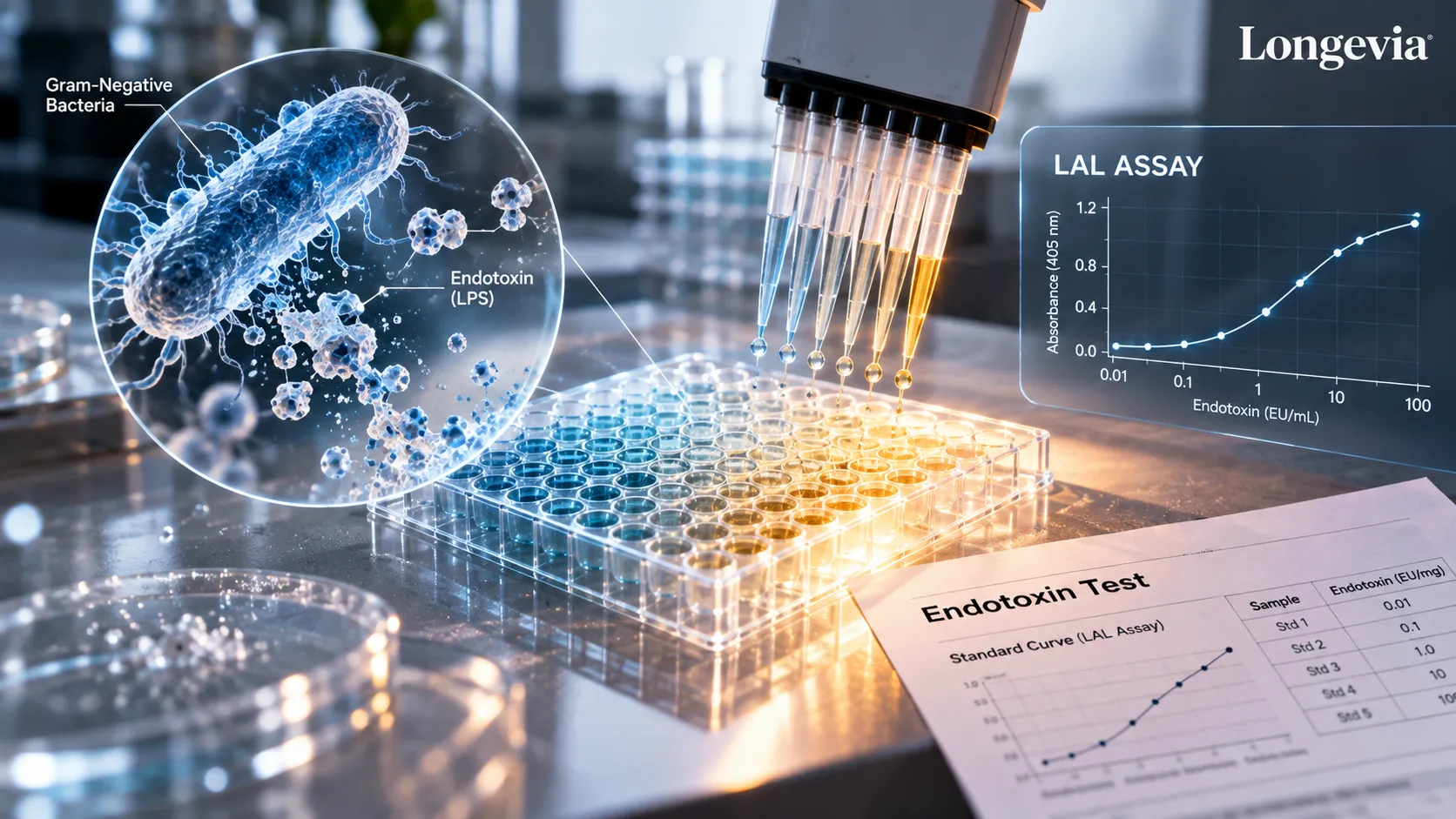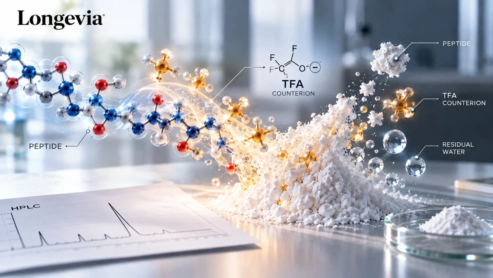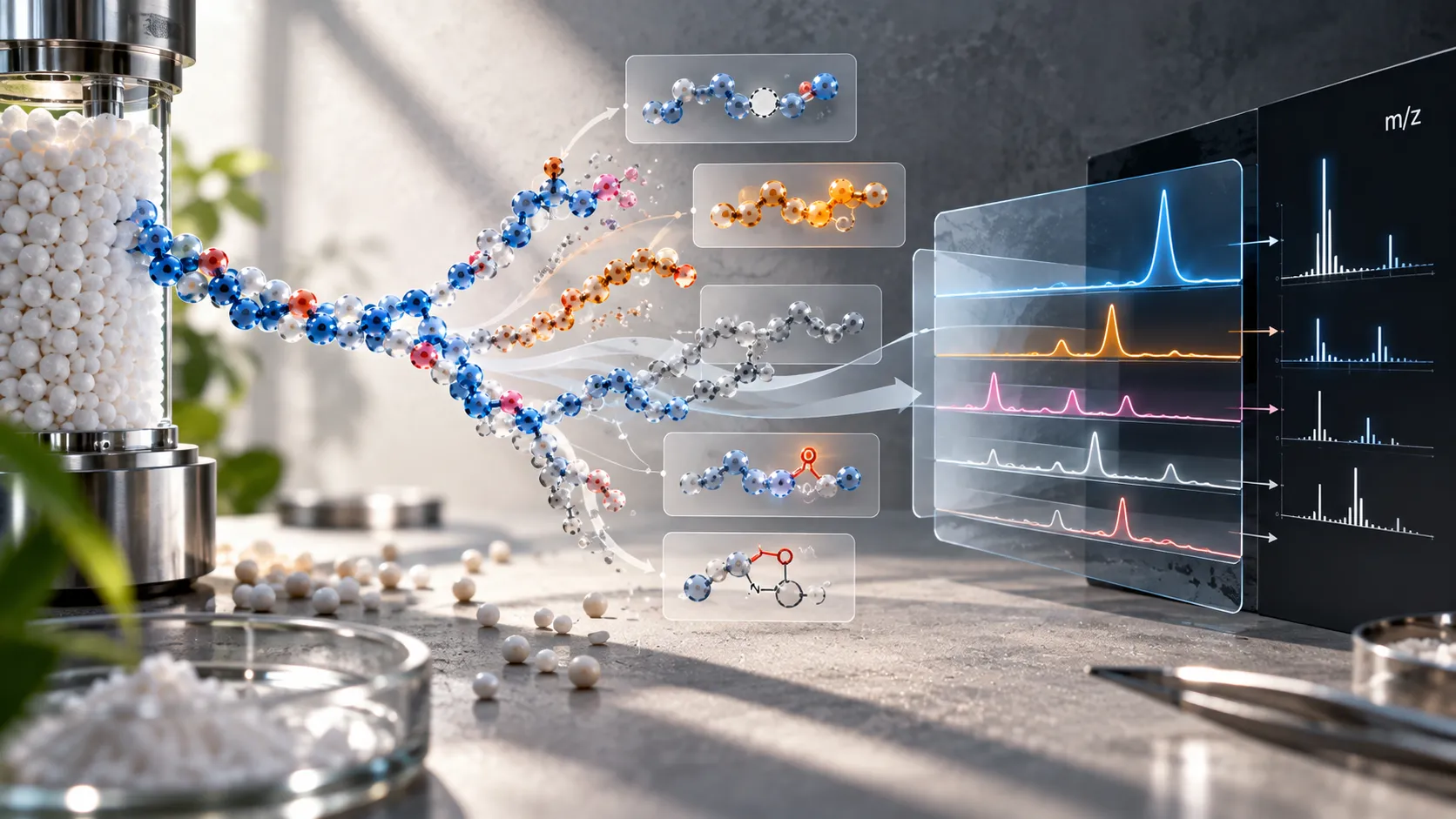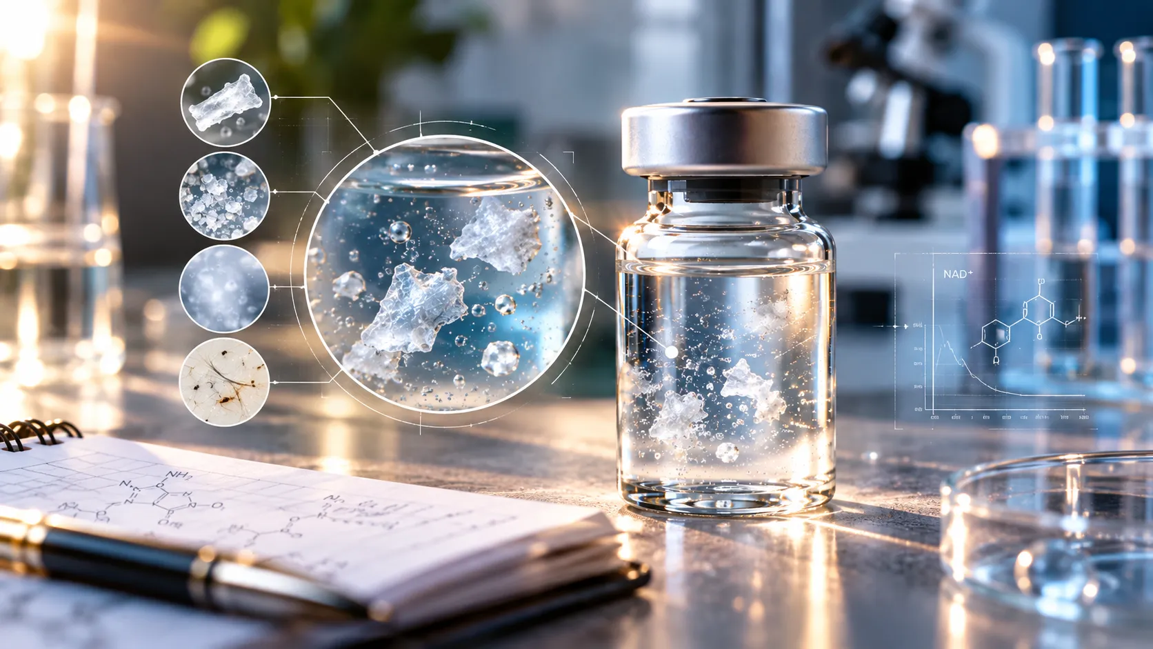
- Four visually distinct appearances point in different directions, and naming which one you have is the first useful step.
- No visual inspection can identify a particle. It narrows the candidates and nothing more.
- Freeze-dried cake that has not fully hydrated can float in the fluid as intact material, and the formulation literature documents this.
- A collapsed or imperfect-looking cake is not a failed product. Controlled studies found reconstitution times unchanged and stability equal or better.
- Glutathione and NAD+ involve different chemistry, and the same explanation should not be applied to both.
- The widely repeated isoelectric point of 5.93 for glutathione has no traceable primary source, and it conflicts with published pKa values.
- Unbuffered NAD+ free acid dissolves to an acidic solution, inside the range suppliers describe as most stable.
- Setting aside a prepared sample is a decision about that sample. It establishes nothing about the source material.
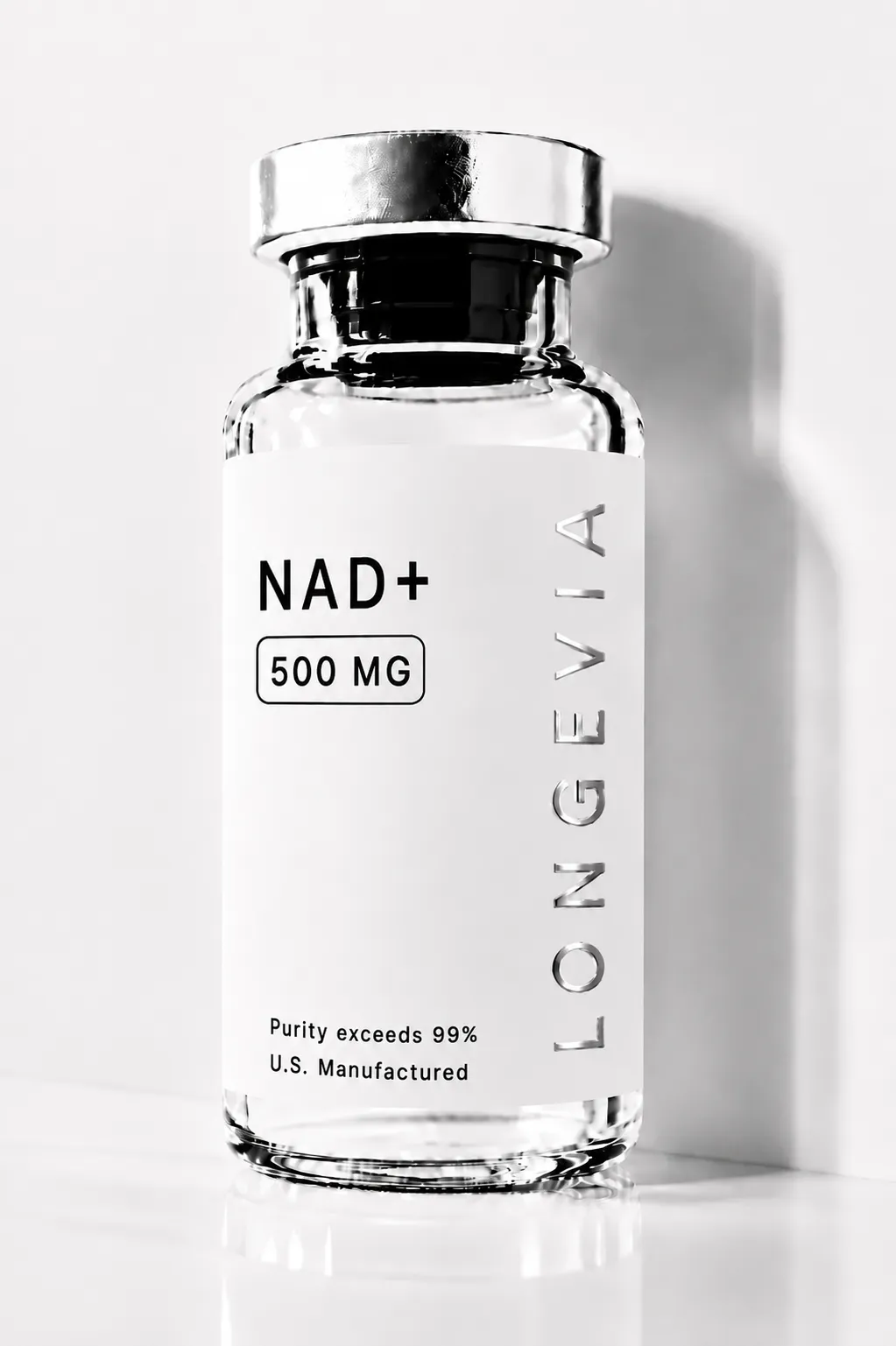
NAD⁺
NAD⁺ (nicotinamide adenine dinucleotide) is a naturally occurring coenzyme investigated in research involving cellular energy metabolism, sirtuin biology, PARP enzyme systems, mitochondrial function, and NAD⁺ homeostasis. Available in 500mg and 1000mg vials. Research Use Only — Longevia Research.
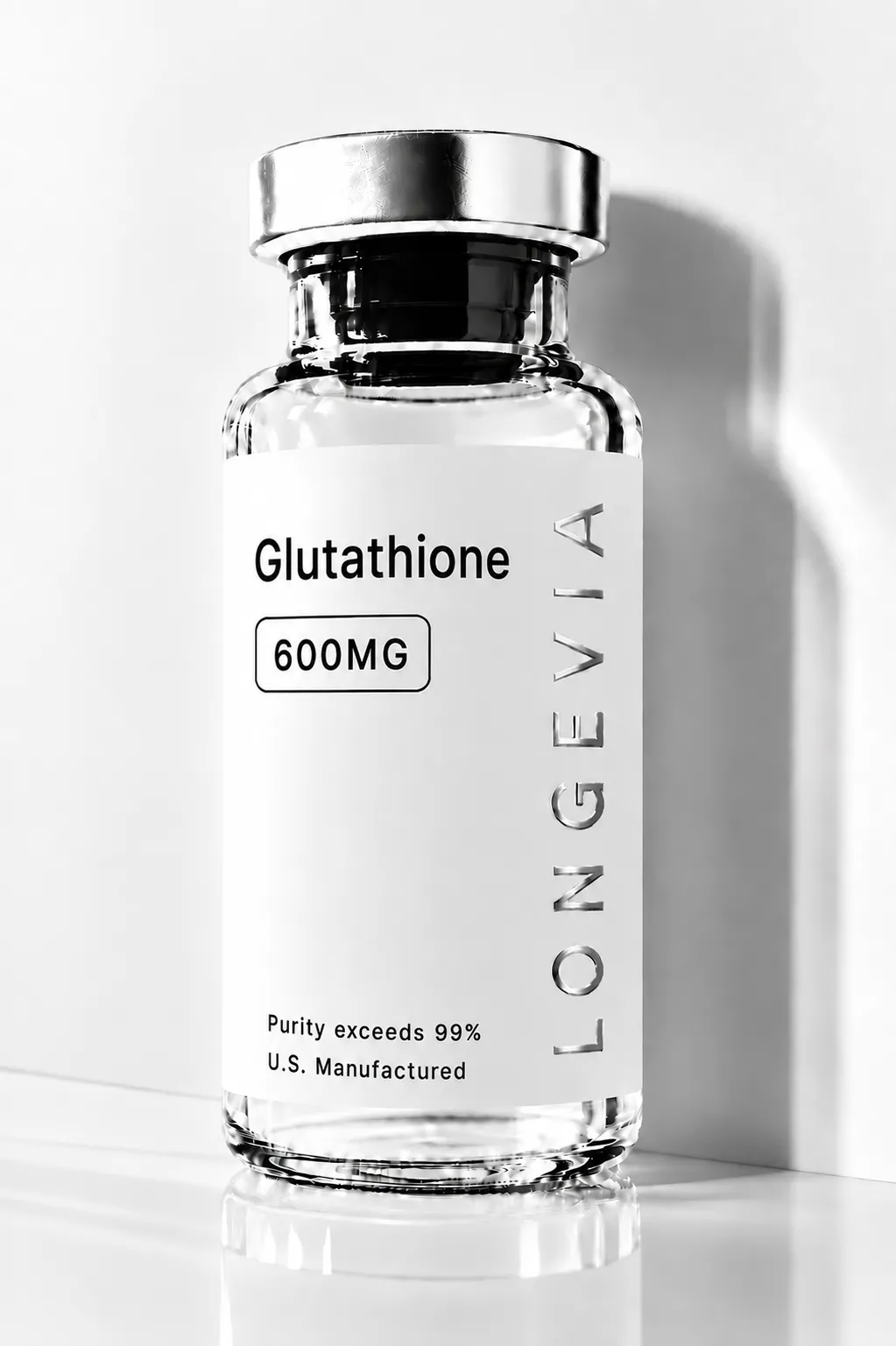
Glutathione
Glutathione (GSH) is a naturally occurring tripeptide antioxidant investigated in research involving oxidative stress biology, glutathione redox cycling, cellular thiol biochemistry, and antioxidant enzyme systems. 600mg and 1500mg vial. Research Use Only — Longevia Research.
NAD⁺
NAD⁺ (nicotinamide adenine dinucleotide) is a naturally occurring coenzyme investigated in research involving cellular energy metabolism, sirtuin biology, PARP enzyme systems, mitochondrial function, and NAD⁺ homeostasis. Available in 500mg and 1000mg vials. Research Use Only — Longevia Research.
Glutathione
Glutathione (GSH) is a naturally occurring tripeptide antioxidant investigated in research involving oxidative stress biology, glutathione redox cycling, cellular thiol biochemistry, and antioxidant enzyme systems. 600mg and 1500mg vial. Research Use Only — Longevia Research.
Visible particles in a reconstituted vial may be undissolved cake material, precipitate, aggregates, degradation-related material, or foreign contamination. Appearance alone cannot establish which of these you have. NAD+ and glutathione fail in different ways, so one explanation does not cover both compounds. What you see should direct the next laboratory question rather than settle it.
Two researchers can prepare the same compound the same way and see different things. One finds a clear solution in under a minute. The other watches a pale mass sit on the surface, apparently refusing to go anywhere. Both observations are common, and neither identifies what is in the vial.
This article covers lyophilized material supplied in vials. Longevia's NAD+ and glutathione sprays ship as ready-to-use liquids and are not reconstituted, so nothing here applies to them.
The aim of this article is deliberately narrow. You will find a way to classify what you are looking at, the mechanisms that could produce it, and what the evidence does not reach. You will not find a preparation procedure or a judgement about whether any preparation can be used.
Research use only. All material described here is for laboratory research use. It is not for human or veterinary use. Nothing here is intended to diagnose, treat, cure, or prevent disease.
Four things researchers actually describe when they say particles
The word covers at least four different observations. They look different and arise from different processes. Lumping them together is how a reader reaches the wrong conclusion about a vial.
What you see | Candidate explanations | Evidence that would separate them | What the observation supports | Appropriate next step |
|---|---|---|---|---|
Translucent or white fragments resembling pieces of the original cake | Incomplete cake hydration; slow dissolution of cake material | Timed re-inspection; comparison with the dry cake; dissolution behaviour under documented handling | Consistent with cake material still dissolving. Consistency is not identification | Record appearance and timing. Treat unexplained persistence as a reason to investigate |
Fine crystalline or sandy material | Precipitation; concentration effects; temperature history | Preparation concentration; temperatures the vial has seen; whether material settles or stays suspended | Consistent with a solubility-related process. Does not establish identity or purity | Record concentration and temperature history first |
Milky haze or gel-like material | Aggregation; a formulation-related problem; a change affecting the whole solution | Whether the haze is uniform; whether it followed a temperature excursion; analytical testing | A change affecting the bulk solution rather than a discrete contaminant | Treat as a hard escalation trigger |
Fibers, dark specks, or other foreign-looking matter | Foreign particulate contamination from the environment, closure, or handling | Shape and colour against contrasting backgrounds; whether a control shows the same | Material that did not originate from the compound | Treat as a hard escalation trigger |
The table narrows the possibilities in front of you. It does not diagnose, and no row should be read as a verdict.
One confusion deserves clearing up separately. Visible bubbles form during reconstitution of freeze-dried material and can persist. Their volume depends on the formulation and on the cake's pore structure. Work on freeze-dried etanercept linked bubble formation to pore size distribution.[1] Work on omalizumab showed that annealing during manufacture sharply reduced bubble volume.[2] Bubbles rise, they are round and uniform, and they are not particles.
What the cake is doing while you wait for it to dissolve
A freeze-dried, or lyophilized, product is not a powder in the ordinary sense. It is a porous solid structure. Water has to travel into that structure before anything dissolves.
The formulation literature describes the sequence in four steps. Fluid wets the cake surface, then penetrates the pores. The wetted cake then disintegrates into pieces. Those pieces finally dissolve, at a rate governed by the viscosity near their surfaces.[3]
What happens when the second step fails is the closest published description of what researchers report seeing. In some amorphous cakes, the fluid did not penetrate the cake at all. An intact cake or a large chunk floated on the fluid among the bubbles, dissolving only by gradual surface erosion.[3] Suspended cake fragments are a documented stage of a normal process, not automatically a defect.
Two intuitions are worth abandoning, because both are common and both are wrong.
The first holds that more surface area means faster dissolution. Researchers studying cooling profiles found longer reconstitution times alongside increased surface area. They attributed the slowdown to closed or small pores blocking water from entering.[4] A study of controlled ice nucleation found the fastest reconstitution at the lowest specific surface area.[5] Pore size and connectivity govern penetration. Total area matters later, once the cake has already broken apart.
Another holds that a collapsed cake means a ruined product. Collapse carries a precise technical meaning here. It is viscous flow of the freeze-concentrated glass above its glass transition temperature. The collapse temperature typically sits one to three degrees above that transition.[6] A controlled study compared collapsed and non-collapsed cakes of identical formulations. It found comparable residual moisture, reconstitution times that were not increased, and no relevant difference in stability.[7] Later work from the same group found stability equal or better in collapsed cakes.[8] Harm from collapse is real in specific systems, and it is not the general rule.
Cake appearance also tells you less than it seems to. Imaging has revealed collapsed internal structure inside lyophilisates that looked pharmaceutically elegant from outside.[6] A cake that looks right has not thereby shown that it is right.
Material quality questions belong to documentation rather than appearance. Longevia's guide to sourcing an NAD+ research compound covers what a Certificate of Analysis does and does not establish.
Glutathione and NAD+ are not the same problem
A tripeptide carrying a reactive thiol and a dinucleotide coenzyme do not fail the same way. Reading across from one to the other reliably produces the wrong explanation.
Glutathione | NAD+ | |
|---|---|---|
Candidate mechanisms | Incomplete hydration; concentration effects; thiol oxidation over time | Incomplete hydration; concentration effects; buffer-specific degradation |
Role of pH | Solution is acidic at the specification concentration. The common pI explanation does not hold | Free acid dissolves acidic. Degradation rates reported independent of pH across pH 4 to 9 |
Concentration | Reported solubility values span roughly tenfold and mostly lack conditions | No documented maximum workable concentration at any stated temperature |
Temperature | Not well characterised in accessible sources for this observation | Not well characterised in accessible sources for this observation |
Cake hydration | Applies, per the general formulation literature | Applies, per the general formulation literature |
Storage-related change | Thiol oxidation is chemically well established | Alkaline lability established. Buffer identity mattered more than pH in one study |
Evidence strength | Analytical specifications and physical chemistry. No product-specific testing | Supplier specifications and a small primary literature. No product-specific testing |
Main open question | What chemical form is supplied, and what is the prepared solution's actual pH | Which salt form is supplied, and at what concentration the material was prepared |
What the evidence supports for glutathione
Start with a figure that carries its conditions. Merck's specification for reduced glutathione, European Pharmacopoeia grade, product 104090, gives pH 3 at 20°C for a 10 g/L aqueous solution.[9] Those stated conditions matter a great deal. A pH value without a concentration and a temperature is not a specification, and it compares with nothing.
Two separate pH explanations circulate for glutathione particles. Neither of them survives close checking.
The original version held that acidic diluent prevents glutathione from dissolving. That runs against the compound's own behaviour. The free acid drives its own solution to pH 3 at a working concentration, dissolving as it does so.
A second version of the story replaced it. In that account, bacteriostatic water near pH 5.7 sits almost exactly on glutathione's isoelectric point of 5.93, where solubility would be lowest. The figure of 5.93 appears widely. It traces to a single chemical-supplier aggregator entry carrying no citation. It also conflicts with the pKa values published on that same page, which are 2.12, 3.53, 8.66 and 9.12 at 25°C. Those values place the zero-net-charge species between the first two, putting the isoelectric region nearer pH 2.8. A measured pH of 3 at 10 g/L fits that picture. No primary, peer-reviewed or pharmacopoeial source for a glutathione isoelectric point could be found, so the 5.93 figure should not be relied on.
The underlying principle needs care too. Textbook organic chemistry states that water solubility is usually lowest at the isoelectric point, and it states this for proteins. No equivalent claim appears for free amino acids or short peptides. Ionic conditions can also move the minimum. One antibody study found its solubility minimum at pH 5.3, well below its isoelectric point of 7.2. Specific anion effects drove that result rather than net charge.[10]
Reported solubility figures conflict badly, and the conflict is instructive. Published values include 50 mg/mL and 61 mg/mL at 25°C from supplier sources, and 100 g/L from the Merck specification.[9][17] Only one of those figures states a temperature. The figures differ roughly tenfold, come from different grades, and do not measure the same quantity. A release specification, a handling figure and an aggregator entry are three different things. No single ceiling can be published responsibly.
Oxidation is better established than any of this. Glutathione's reactive species is the thiolate anion rather than the protonated thiol. Primary kinetic work reports the thiol pKa at 8.8 to 8.94.[11][12] Reactivity rises steeply from pH 7 toward pH 9, then levels off near the pKa. There is no threshold at pH 7. Only a small percentage exists as thiolate at pH 7, so the shift is a curve rather than a switch.
One further variable applies to synthetic peptide material rather than to either compound here. A residual counterion left from synthesis adds mass and can shift a prepared solution's acidity, as Longevia's guide to TFA counterions and residual water explains. Glutathione supplied as the free acid carries no such counterion, and the product page does not state which form it supplies.
Compound background and the limits of reconstitution-related claims appear in Longevia's article on glutathione and skin research.
What the evidence supports for NAD+
The most useful verified fact about NAD+ runs opposite to what many readers expect. Glentham's specification for NAD+ free acid gives pH 2.0 to 4.0 for a 10% aqueous solution.[18] Unbuffered NAD+ free acid dissolves to an acidic solution.
Supplier stability guidance points the same way. Sigma's product information for beta-NAD free acid recommends aqueous solutions between pH 2 and 6 for storage.[19] It describes the compound as very labile in alkaline solution, particularly alongside phosphate, maleate or carbonate. That sheet also mentions adjusting the pH to 7 with sodium hydroxide. It offers this as a route into solution, not as a stability target.
Claims that NAD+ stability is straightforwardly pH dependent need qualifying. Wong and Whitesides reported decomposition rates for oxidized nicotinamide cofactors as effectively independent of pH from 4 to 9. Instability appeared at the extremes and alongside strong nucleophiles. A recent study reports an optimum near pH 8.5 for the NAD+ and NADH system together. That figure reaches this article second-hand, through the paper's citation of earlier work.[13]
That same study found something more useful for anyone preparing solutions. At a fixed pH, buffer identity mattered more than anything else. NAD+ in Tris lost very little over 43 days, while NAD+ in HEPES degraded almost entirely.[13] The authors describe their NAD+ analysis as qualitative, so no rate should be taken from it.
Concentration deserves a plain statement of the position. No authoritative maximum workable aqueous concentration for NAD+ has been documented at any stated temperature. Supplier solubility figures of 50 mg/mL and 33.3 mg/mL appear without temperatures[19] and read as test thresholds rather than saturation limits. Arithmetic involving high concentrations, such as 500 mg in 3 mL giving roughly 167 mg/mL, is arithmetic. It carries no recommendation and sets no limit.
Yellowing deserves much the same treatment. Faint yellow is within specification for fresh NAD+ material. Supplier descriptions cover white to light yellow powder, and a solution described as colourless to faintly yellow.[18][19] Colour therefore cannot by itself signal degradation. No primary source links visible discoloration of NAD+ solutions to a specific degradation pathway, and inferring one from appearance is not supportable.
One limit applies to everything above. Longevia's NAD+ product page gives free-acid identifiers and directs researchers to the batch Certificate of Analysis. Salt form and hydration change the molecular weight. Until that form is confirmed for a lot, no pH statement here applies to a specific vial.
Particles on the first day and particles after a week
Timing narrows the candidates a little. It settles nothing on its own, and assigning a mechanism from the timeline alone is a mistake.
Material visible immediately after preparation is consistent with incomplete cake hydration, undissolved solids, concentration effects, or preparation temperature. These are physical processes, and the cake section above covers the best-documented one.
Something appearing in a previously clear solution raises different questions. Chemical change, oxidation, temperature history and contamination introduced on re-entry all belong here. Glutathione oxidation and NAD+ degradation are separate chemical questions with separate evidence. Neither should be used to explain the other.
Storage can also change the solid before anything is added to it. Moisture can diffuse into dried material from the closure, lowering the temperature at which the solid collapses.[14] Water acts as a plasticiser, so higher residual moisture lowers the cake's glass transition.[15] Whether such a cake then reconstitutes measurably more slowly is an inference from that chain, not a measured finding.
Storage conditions and timelines for prepared solutions appear in Longevia's guide to how long peptides last after reconstitution.
What the diluent can and cannot change
Longevia's Reconstitution Solution is water for injection containing 0.9% benzyl alcohol as a preservative. The product page states no pH.
A figure of pH 5.7, with a range of 4.5 to 7.0, appears in Longevia's article on bacteriostatic water and sterile water. That figure carries no inline citation there. Treat it as a reference value for a class of product, not a measurement of any lot.
More importantly, a diluent's nominal pH does not establish the pH of the solution after a compound dissolves in it. Bacteriostatic water is unbuffered, and both compounds here have their own acid-base behaviour. Predicting a final pH is not possible from the available information, and this article prescribes no diluent, volume or ratio.
A proposed way to assess a questionable preparation
What follows records and classifies an observation. It is not a validated method, and no validated protocol for this assessment exists. It produces documentation rather than a verdict. Nothing in it is intended to make a questionable preparation look acceptable.
- Describe the appearance against a matt dark background, then against a plain light one. Note colour, whether material is suspended or settled, and whether it is uniform or discrete.
- Record when it appeared relative to preparation, and whether the solution was ever clear.
- Record the preparation concentration, the diluent used, and every temperature the vial has experienced.
- Note the lot number, and whether that lot's Certificate of Analysis is available.
- Re-inspect after an interval and record any change, without altering the preparation in any way.
Nothing in that sequence involves warming, adding diluent, filtering, or agitating beyond what product documentation supports. No step is aimed at clearing the solution.
Certain observations should end the assessment and move to escalation. Fibers or foreign-looking specks belong here. So do persistent milky haze or gel-like material, unexplained discoloration, and particles appearing after a previously clear solution was stored. Anything unexplained after careful inspection belongs here too, as does any missing identity or quality documentation.
Setting aside a prepared sample is a decision about that sample. It does not establish that the source material was contaminated, misidentified or poorly manufactured, and it should not be reported as though it did.
What none of this can establish
The specific gaps deserve naming rather than gesturing at.
Neither Longevia product page states the salt form supplied. The house diluent discloses no pH. No mechanism described here has been tested on either product, and nothing in this article reports a measurement made on Longevia material. Visual observation cannot distinguish a precipitate from a degradation product from a contaminant.
Pharmacopoeial standards are narrower than they sound. European Pharmacopoeia requirements for a freeze-dried product apply to the reconstituted solution rather than the dry cake. The phrases "essentially free" and "practically free" of visible particles describe a probabilistic standard depending on defined inspection conditions.[16] They do not mean an absence of particles. These are release specifications for commercial parenteral products, inspected under controlled lighting and defined viewing times. They establish nothing for research-use material.
Prevention, within those limits, comes down to documentation. Keep each lot's Certificate of Analysis and know what it reports, requesting it where none is published. Record preparation details at the time rather than reconstructing them later. Treat anything you cannot explain as a reason to investigate rather than proceed.
Two measurements would resolve most of what is open. Confirming the chemical form supplied does the first part. Measuring the pH of a prepared solution at a stated concentration does the rest.
What to write down before you do anything else
Appearance narrows a question and never answers it. Four visually distinct observations point in different directions. Cake hydration explains more of what researchers see than solution pH does, and the two compounds fail by different routes.
A written description of what you saw, when it appeared, and which lot it came from is what makes later investigation possible. It costs nothing and it is always available.
Longevia publishes lot-specific Certificates of Analysis on its certificates page, where identity and purity questions for a given lot belong. Coverage there is partial, so request the document for your lot if it is not listed.
Literature retrieved via PubMed.
Frequently Asked Questions
- Gao H, Du CY, Zheng A, Qian C, Fang WJ. Formulation Factors Affecting the Formation of Visible-Bubbles During the Reconstitution Process of Freeze-Dried Etanercept Formulations. AAPS J. 2025;27(1):29.
- Gao H, Ge XZ, Liu JW, Wang ST, Xu J, Fang WJ. Effect of Annealing on Visible-Bubble Formation and Stability Profiles of Freeze-Dried High Concentration Omalizumab Formulations. Mol Pharm. 2024;21(4):1691-1704.
- Key factors governing the reconstitution time of high concentration lyophilized protein formulations. Eur J Pharm Biopharm. 2021;165:361-373.
- Insights into the influence of the cooling profile on the reconstitution times of amorphous lyophilized protein formulations. Eur J Pharm Biopharm. 2015;96:247-254.
- Wang J, Searles JA, Torres E, Tchessalov SA, Young AL. Impact of Annealing and Controlled Ice Nucleation on Properties of A Lyophilized 50 mg/ml MAB Formulation. J Pharm Sci. 2022;111(9):2639-2644.
- Haeuser C, Goldbach P, Huwyler J, Friess W, Allmendinger A. Be Aggressive! Amorphous Excipients Enabling Single-Step Freeze-Drying of Monoclonal Antibody Formulations. Pharmaceutics. 2019;11(11):616.
- Schersch K, Betz O, Garidel P, Muehlau S, Bassarab S, Winter G. Systematic investigation of the effect of lyophilizate collapse on pharmaceutically relevant proteins I. J Pharm Sci. 2010;99(5):2256-2278.
- Schersch K, Betz O, Garidel P, et al. Systematic investigation of the effect of lyophilizate collapse on pharmaceutically relevant proteins, part 2. J Pharm Sci. 2012;101(7):2288-2306.
- Merck/Sigma-Aldrich. Glutathione (reduced) EMPROVE EXPERT Ph Eur, product 104090. Product specification.
- Zhang L, Tan H, Fesinmeyer RM, et al. Antibody solubility behavior in monovalent salt solutions reveals specific anion effects at low ionic strength. J Pharm Sci. 2012;101(3):965-977.
- Baker LMS, Baker PRS, Golin-Bisello F, et al. Nitro-fatty acid reaction with glutathione and cysteine. J Biol Chem. 2007;282(42):31085-31093.
- Turell L, Vitturi DA, Coitiño EL, et al. The Chemical Basis of Thiol Addition to Nitro-conjugated Linoleic Acid. J Biol Chem. 2017;292(4):1145-1159.
- Wolfe KD, Alahuhta M, Himmel ME, Bomble YJ, Jennings GK, Cliffel DE. Long-Term Stability of Nicotinamide Cofactors in Common Aqueous Buffers. Molecules. 2024;29(22):5453.
- Adams GD, Ramsay JR. Optimizing the lyophilization cycle and the consequences of collapse on the pharmaceutical acceptability of Erwinia L-asparaginase. J Pharm Sci. 1996;85(12):1301-1305.
- te Booy MP, de Ruiter RA, de Meere AL. Evaluation of the physical stability of freeze-dried sucrose-containing formulations by differential scanning calorimetry. Pharm Res. 1992;9(1):109-114.
- Mathonet S, Mahler HC, Esswein ST, et al. A Biopharmaceutical Industry Perspective on the Control of Visible Particles in Biotechnology-Derived Injectable Drug Products. PDA J Pharm Sci Technol. 2016;70(4):392-408.
- Selleck Chemicals. GSH (L-Glutathione reduced), catalog S4606. Product datasheet.
- Glentham Life Sciences. beta-Nicotinamide adenine dinucleotide, product GV0860. Product specification.
- Sigma-Aldrich. beta-Nicotinamide adenine dinucleotide, free acid, product N8410. Product information sheet.