- Endotoxin is lipopolysaccharide from the outer membrane of Gram-negative bacteria, and lipid A is the biologically active part.
- A COA with no endotoxin line means the test was not reported, not that the lot passed.
- Sterility and endotoxin are different tests, and a sterile sample can still contain endotoxin.
- Endotoxin units measure biological activity against a reference standard, not mass.
- An EU/mg figure shifts depending on whether the milligram is gross powder or net peptide.
- Cultured monocytes and macrophages respond to trace endotoxin through TLR4, which can confound cytokine read-outs.
- USP chapter 85 requires a spiked interference check, with recovery between 50 and 200 percent.
- Recombinant factor C assays detect endotoxin without horseshoe crab lysate and are covered by Ph. Eur. 2.6.32 and USP chapter 86.
Autoclaving kills the bacteria in a solution. It does not reliably destroy the endotoxin they leave behind. That material is heat stable. Depyrogenating glassware calls for dry heat near 250 degrees Celsius, held for about 30 minutes. A 0.22 micron filter does not stop it either. So endotoxin testing peptides is a separate question from sterility. The two tests answer different things. This guide covers what the test measures and how the LAL assay works. It also covers what EU and EU/mg mean. Then it looks at why background endotoxin wrecks a cell-based read-out. Last, what to ask when a COA carries no endotoxin line.
An endotoxin test measures lipopolysaccharide from the outer wall of Gram-negative bacteria. The LAL assay is the usual method, and the result is reported in endotoxin units. It is separate from sterility. If a COA lists no endotoxin result, the test was not reported.
What is endotoxin and where does it come from?
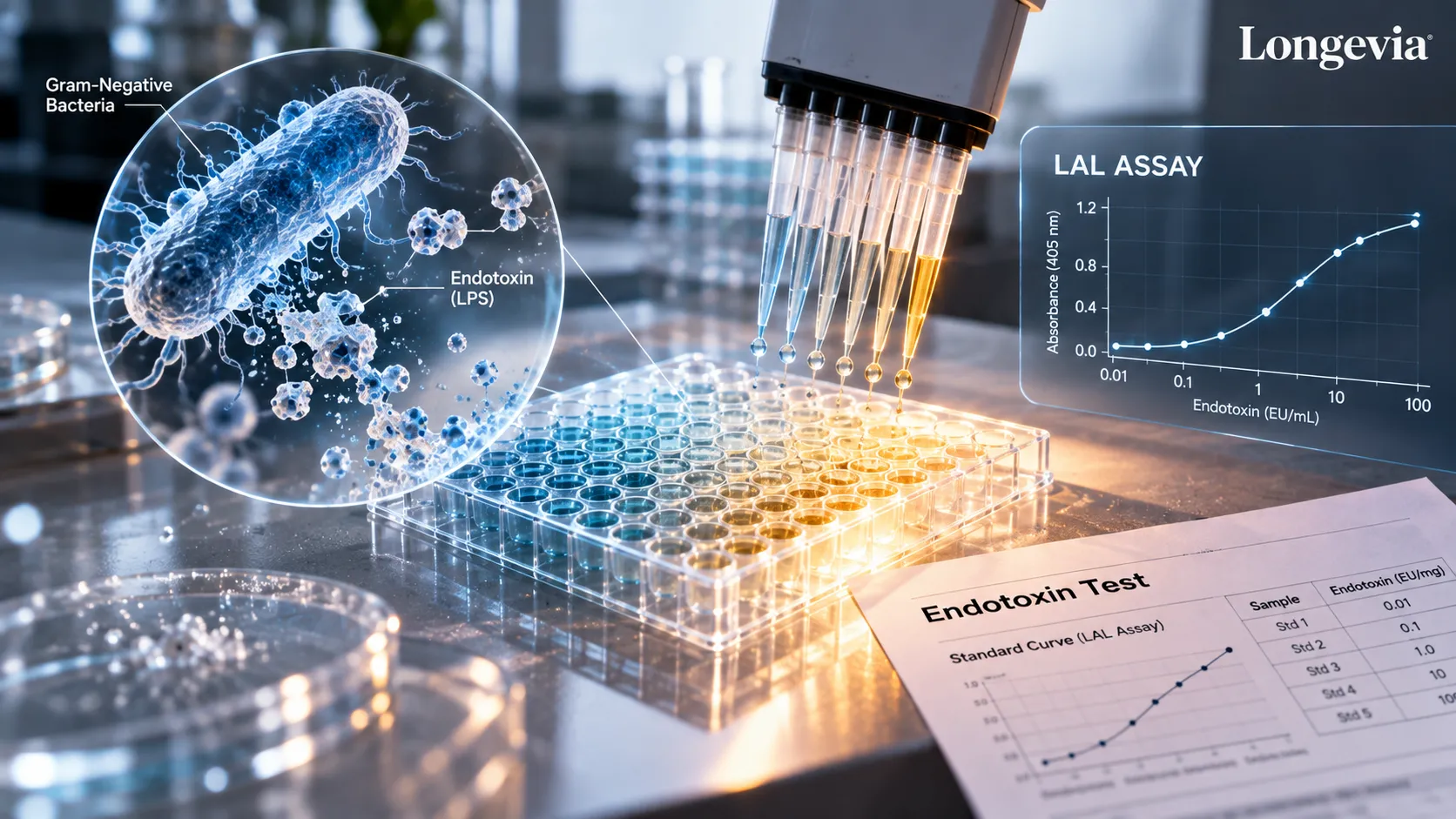
Endotoxin is lipopolysaccharide, or LPS, a large molecule built into the outer membrane of Gram-negative bacteria. The biologically active part is lipid A, the fatty anchor at one end.
Bacteria shed LPS as they grow. They release more when they die. Killing the cells does not remove what they leave behind. Heat makes the point well. Standard autoclave cycles sterilize without dependably destroying endotoxin. Compendial depyrogenation of glassware uses dry heat instead, commonly 250 degrees Celsius for 30 minutes.
Filtration leaves the same gap. A 0.22 micron membrane holds back whole bacteria. LPS molecules and their aggregates pass straight through into the filtrate.
Manufacturing adds its own openings. Synthesis, cleavage, purification, lyophilization, and vial filling each involve solvents, resins, and equipment. Any one of those steps can introduce contamination or leave existing contamination in place. Nothing in a standard purification sequence targets LPS specifically.
Gram-positive bacteria are a different case. They have no outer membrane, so they carry no LPS. They can still shed other pyrogenic material. That distinction matters when you read a test that looks for endotoxin and nothing else.
Contamination routes in a laboratory are mundane. Water is the usual one, because Gram-negative organisms colonize purified water systems happily. Buffers, salts, culture media, glassware, plastic consumables, and raw starting materials each add a share. So does handling. Pipette tips, gloves, and open tubes collect whatever sits on the bench.
Surfaces matter as well. Gorbet and Sefton reviewed biomaterials work in a 2005 review. They describe how strongly endotoxin adsorbs and how hard it is to exclude from normal practice. For a solid product, the contaminant can arrive on the glass or in the powder rather than in the solvent.
Quantity is the part people underestimate. Cultured immune cells respond to picogram amounts. That sits far below anything visible in a vial. It also sits far below what a purity chromatogram would flag. Purity and endotoxin answer different questions, and our guide to HPLC purity on a COA covers what that number describes.
How does the LAL test work?
The LAL test uses a clotting cascade taken from horseshoe crab blood cells. It fires when it meets endotoxin. What the instrument records is how fast, or how far, that cascade runs.
Limulus amebocyte lysate comes from amebocytes, the blood cells of the horseshoe crab. Contact with LPS activates factor C, the first enzyme in the chain. Downstream enzymes convert a clotting protein and the mixture gels. Three read-outs of that reaction are in routine use. The harmonized pharmacopoeial chapters treat them as equivalent methods.
Method | What you read | Quantitative or limit | Note |
|---|---|---|---|
Gel-clot | Whether a firm gel forms in the tube | Limit test, or semi-quantitative by dilution | Referee method under USP 85 when results are disputed |
Turbidimetric LAL | Cloudiness developing over time | Quantitative, against a standard curve | Kinetic and end-point versions both exist |
Chromogenic LAL | Color released from a synthetic peptide substrate | Quantitative, against a standard curve | Tolerates turbid samples better than gel-clot |
Recombinant factor C | Fluorescence, or color in some formats | Quantitative, against a standard curve | No crab lysate. Ph. Eur. 2.6.32 and USP 86 |
Sensitivity comes from the reagent, not from the method name. Gel-clot lysate carries a labeled sensitivity, written as lambda. The test then answers only above or below that level. Photometric methods report a value against a standard curve, with a reporting limit of their own. A number means little without that limit beside it.
Dilution has a ceiling as well. The maximum valid dilution is the furthest you can dilute a sample and still detect the limit you care about. Push past it and a clean result stops meaning anything. Interference is often fixed by dilution, so the two constraints pull against each other.
Recombinant factor C drops the animal-derived lysate entirely. Ding and Ho set out the case for it in a 2001 review. Their argument rests on one finding. Factor C alone is the endotoxin sensor in the cascade. Ph. Eur. chapter 2.6.32 describes the method, and USP added chapter 86 for recombinant reagents, which took effect in 2025.
Specificity is the other practical advantage. rFC does not react to beta-glucans. Those can trip an LAL result and produce a false positive that has nothing to do with bacteria.
A different route exists for pyrogens more broadly. The monocyte activation test is Ph. Eur. chapter 2.6.30. It incubates human monocytes with the sample and measures cytokine release. That makes it sensitive to fever-causing material which is not endotoxin.
What do EU, EU/mL, and EU/mg mean?
An endotoxin unit, written EU, is a unit of biological activity rather than mass. It is defined against a reference standard endotoxin. One EU means one fixed amount of reactivity in the assay.
Activity is the right basis because mass would mislead. LPS molecular weights range enormously. Potency also shifts with the bacterial species, the acylation pattern of lipid A, and how the material aggregates. Sources commonly put 1 EU at roughly 100 picograms of E. coli LPS. Treat that as an order-of-magnitude guide, not a conversion factor.
Units follow the form of the sample. Liquids are reported in EU/mL, endotoxin units per milliliter. Solids are reported in EU/mg, endotoxin units per milligram. The laboratory dissolves a weighed amount, reads the solution, then converts back to a mass basis.
That conversion is where per-mg figures get slippery. A milligram of powder is not a milligram of peptide. Lyophilized material also carries counterion, residual water, and salts. A figure quoted per milligram gross looks lower than the same figure per milligram of net peptide. Our post on net peptide content and HPLC purity explains why the two bases diverge.
Here is a worked example using invented round numbers. Say a laboratory reports 0.50 EU per milligram of powder. Say the same lot is 80 percent net peptide by mass. Divide 0.50 by 0.80 and you get about 0.63 EU per milligram of peptide. One lot, one measurement, two numbers a quarter apart.
Read a less-than result carefully too. A report of less than 0.10 EU/mg says the assay found nothing down to its limit. It does not say the sample is free of endotoxin. A more sensitive method might have found something under that line.
How is endotoxin different from sterility and bioburden?
Sterility asks whether anything living grows. Endotoxin asks whether a specific bacterial molecule is present. A sample can pass one and fail the other, in either direction.
Property | What it means | How it is tested | Independent of the others |
|---|---|---|---|
Endotoxin | LPS from Gram-negative cell walls, live or dead cells | LAL or recombinant factor C, USP 85 and 86 | Yes. A sterile sample can still contain endotoxin |
Sterility | No viable organisms grow under defined conditions | Growth-based culture, USP 71 | Yes. Sterile says nothing about residue left behind |
Bioburden | The count of viable organisms present | Plate counts and enumeration, USP 61 | Yes. A low count does not imply low endotoxin |
Pyrogenicity | Capacity to cause a fever response in a test system | Rabbit pyrogen test USP 151, or monocyte activation test Ph. Eur. 2.6.30 | Yes. Some pyrogens are not endotoxin |
Worth stating flatly: a sterile sample can still contain endotoxin. Sterilization kills organisms. It does not remove the molecules they leave behind. That gap is why USP keeps chapter 71 for sterility separate from chapter 85 for bacterial endotoxins.
The reverse holds too. A powder with a measurable bioburden may show little endotoxin. That happens when the organisms present are not Gram-negative. Neither result predicts the other well enough to stand in for it.
Most research peptide COAs report none of these four attributes. Identity by mass spectrometry, purity by HPLC, appearance, and net content are the usual set. Microbiological attributes sit outside that scope. Knowing this before you go looking saves a round of email.
Why can endotoxin ruin a cell-based assay?
The evidence on this point comes from in-vitro cell-culture work. Monocytes and macrophages detect LPS through the TLR4 and MD-2 receptor complex. They respond at very low concentrations by switching on inflammatory signaling.
That sensitivity is the problem. Background endotoxin in a reagent produces the same read-out a researcher might credit to the test compound. Gorbet and Sefton, reviewing biomaterials studies, found repeated cases of exactly this. Contaminated preparations induced cytokine production that vanished once the endotoxin was removed.
A cytokine assay shows the effect most directly. Measure TNF-alpha or IL-6 release from monocytes. Add a reagent carrying trace LPS. The plate now reports a response with no connection to the molecule under study.
Media components carry their own risk. Serum, growth factors, and supplements each pass through manufacturing steps where contamination can happen. Swapping a serum lot mid-experiment can shift baseline cytokine output on its own.
Two controls settle most arguments quickly. One is a matched vehicle prepared from the same reagents as the treatment. The other is an endotoxin-binding agent such as polymyxin B, run in parallel. If the signal drops when endotoxin is blocked, you have your answer.
Not every assay is this fragile. Sensitivity depends on the cell type, the read-out, and whether the measured pathway intersects TLR4 signaling. A binding assay with purified protein will usually not care. A chemical stability study will not care either. Primary immune cells, whole blood work, and inflammatory read-outs care a great deal.
Why do peptide COAs rarely list endotoxin?
Endotoxin on a certificate of analysis is the exception rather than the rule for research peptides. Three practical reasons explain that. None of them is a statement about the material itself.
Cost is the first. An endotoxin test is a separate assay with its own reagents and controls. It also needs method suitability work for each product. The whole package is priced per lot.
The second reason is scope. Pharmacopoeial endotoxin limits are written for pharmaceutical products. They are calculated from how those products are used. They are not a statement about research material. A reagent sold for in-vitro work sits outside that framework entirely.
Demand explains the rest. Many buyers run assays where trace endotoxin does not change the result. Suppliers test for what most customers need. Identity, purity, and mass confirmation cover the common cases. Our post on why peptide purity matters covers what those routine tests settle and what they leave open.
None of this says anything about a specific lot. A supplier that prints no endotoxin figure may still hold clean material. A supplier that prints one may have tested under conditions unlike your assay. The line on the page records what was measured, nothing more.
A COA with no endotoxin line means the test was not reported. It does not mean the lot passed. It does not mean the lot failed. Absence of a result is absence of information.
Can a peptide interfere with the LAL test itself?
Yes, samples can inhibit or enhance the reaction. That is why the method requires an interference check before any result counts. The check spikes a known amount of endotoxin into the sample. Then it asks whether the assay finds that spike again.
USP chapter 85 sets the acceptance window at 50 to 200 percent of the added amount. That figure is worked out after subtracting whatever the unspiked sample shows. Recovery below 50 percent points to inhibition. Recovery above 200 percent points to enhancement. Either way the method is unsuitable until the interference is dealt with, usually by dilution.
Low endotoxin recovery is a related but distinct problem. It is well documented in the analytical literature. In certain matrices, spiked endotoxin becomes undetectable over hours or days. The usual controls still look fine throughout. Chelators combined with surfactants are the classic trigger. Schwarz and colleagues showed in a 2017 analytical and cell-culture study that masked endotoxin stays biologically active. Dilution does not reverse the masking.
Charge and shape matter for peptides specifically. Cationic and amphipathic peptides bind lipid A through its anionic phosphate groups. Several have been shown to suppress the LAL signal. David and colleagues reported this in a 2000 analytical study. LPS-binding peptides inhibited both gel-clot and chromogenic read-outs, to differing degrees.
The consequence is worth spelling out. A peptide with that character can change what the assay sees. It does not change what is actually in the vial. Only the interference check separates those two situations.
False positives run the other way. LAL reagent also responds to beta-glucans from cellulose filters and some plant-derived materials. A blocking buffer or an rFC method removes that route.
What should I ask for if my assay is endotoxin-sensitive?
Ask for the number and the context around it. A bare figure with no method, no limit, and no lot number is not something you can act on. Suppliers of low endotoxin research reagents usually have these details to hand.
What to ask | Why it matters |
|---|---|
Which method, gel-clot, kinetic chromogenic, or recombinant factor C | Limit tests and quantitative tests answer different questions |
The assay sensitivity or reporting limit | A "less than" result only means something next to the limit |
Whether the result is lot specific | A generic figure does not describe the vial in your hand |
The unit and the basis, per mg gross powder or per mg net peptide | One lot yields two different numbers on the two bases |
Whether interference was checked with a spiked control | Recovery outside 50 to 200 percent invalidates the reading |
Test date and the laboratory that ran it | Lets you trace the result and judge how current it is |
Housekeeping in your own laboratory does the rest. Use endotoxin-free water, buffers, and plasticware when setting up assays. Keep a dedicated set of consumables for endotoxin-sensitive work. Depyrogenate glassware by dry heat rather than assuming an autoclave cycle handled it.
Controls close the gap a missing COA line leaves open. Run a vehicle control prepared from the same water and buffers as the test condition. If the vehicle produces signal, your reagents are the suspect.
Testing in house is more approachable than it used to be. Kinetic chromogenic and rFC kits run on a standard microplate reader. One plate can screen several lots at once. You also keep the record yourself, which is worth something when a reviewer asks.
Write down what you learn. Record the method, the limit, the lot number, and the test date alongside your own results. Reviewers ask about reagent provenance more often than they used to. A short note now saves rebuilding the trail later.
Endotoxin-free water, buffers, and plasticware cost little next to a repeated experiment. Dedicate a set for endotoxin-sensitive assays and label it clearly.
What is a fair way to handle a COA with no endotoxin line?
Three steps, in order of effort.
- Ask the supplier. Request the method, the limit, the lot-specific result, and whether interference was checked. A clear no tells you more than a vague yes.
- Test it yourself. Commercial LAL and rFC kits run on a standard plate reader. Testing the lot in your hands removes the guesswork.
- Add controls. Where testing is not practical, use a vehicle control and a known-clean comparator. Those will show whether background signal is present.
Longevia Research has every batch independently tested by HPLC and LC-MS. Lot-specific COAs are published in the COA Library.
For laboratory research use only. Not for human or veterinary use. Not intended to diagnose, treat, cure, or prevent disease.
Frequently Asked Questions
- United States Pharmacopeia. General Chapter 85, Bacterial Endotoxins Test (pharmacopoeial standard, harmonized with Ph. Eur. 2.6.14 and JP 4.01).
- United States Pharmacopeia. General Chapter 86, Bacterial Endotoxins Test Using Recombinant Reagents (pharmacopoeial standard).
- US Food and Drug Administration. Pyrogen and Endotoxins Testing: Questions and Answers, Guidance for Industry, March 2026 (regulatory document).
- Gorbet MB, Sefton MV. Endotoxin: the uninvited guest. Biomaterials. 2005;26(34):6811-6817 (review).
- Ding JL, Ho B. A new era in pyrogen testing. Trends in Biotechnology. 2001;19(8):277-281 (review).
- Schwarz H, Gornicec J, Neuper T, et al. Biological activity of masked endotoxin. Scientific Reports. 2017;7:44750 (analytical and cell-culture study).
- David SA, Awasthi SK, Balaram P. The role of polar and facial amphipathic character in determining lipopolysaccharide-binding properties in synthetic cationic peptides. Journal of Endotoxin Research. 2000;6(3):249-256 (analytical-method study).