- Reconstitution concentration equals peptide mass divided by diluent volume, so 10 mg of MOTS-c in 2 mL yields 5 mg/mL.
- MOTS-c is a 16-amino-acid mitochondrial-derived peptide with the sequence MRWQEMGYIFYPRKLR and an average molecular weight of approximately 2,175 Da.
- Net peptide content is lower than gross vial mass because of counterion and residual water, so calculations from the label value overstate concentration.
- Purity by HPLC and net peptide content are different parameters and a certificate of analysis should report both.
- The MOTS-c sequence contains two methionines and one tryptophan, making oxidation the primary chemical degradation risk.
- MOTS-c contains no cysteine, so disulfide scrambling and disulfide-mediated aggregation are not available degradation pathways.
- Reconstituted peptide requires refrigeration and light protection, and repeated freeze-thaw cycling should be avoided by aliquoting before freezing.
- FDA's Pharmacy Compounding Advisory Committee reviewed MOTS-c-related bulk drug substances in July 2026, and MOTS-c is not an FDA-approved drug for any indication.
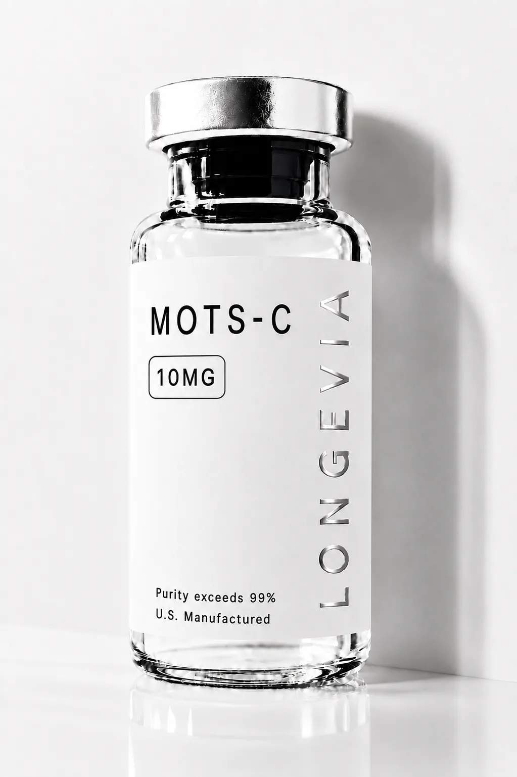
MOTS-C
Buy research-grade MOTS-C with COA-verified 99%+ purity. HPLC-tested, US-based, lab-quality mitochondrial peptide for research use only.
MOTS-C
Buy research-grade MOTS-C with COA-verified 99%+ purity. HPLC-tested, US-based, lab-quality mitochondrial peptide for research use only.
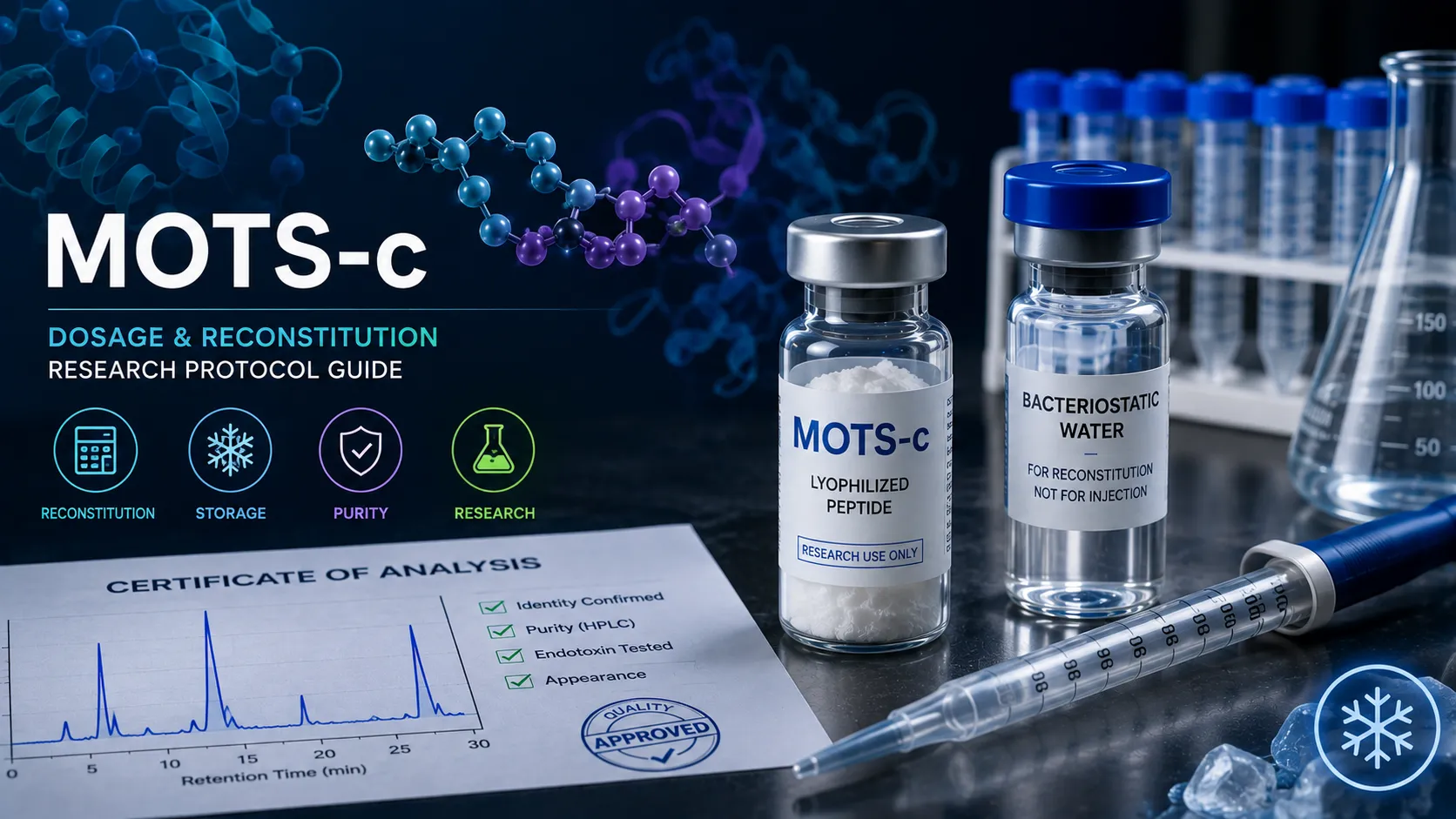
MOTS-c is a 16-amino-acid mitochondrial-derived peptide, and most handling errors researchers make with it trace back to two places: the arithmetic that converts vial mass into working concentration, and the storage decisions made in the first few minutes after diluent goes into the vial. This guide covers reconstitution math, diluent selection, storage conditions, and the degradation risks that follow directly from this peptide's amino acid composition. It is an operational handling reference rather than a mechanism review, so the biology is summarised briefly and the depth sits in the protocol. This material is supplied for research use only (RUO), is not for human consumption, and carries no therapeutic or diagnostic claim.
Reconstitution concentration is vial mass divided by diluent volume. Adding 2 mL of bacteriostatic water to a 10 mg vial yields 5 mg/mL, which is 500 mcg per 0.1 mL. Diluent volume is the only variable under the researcher's control at this step, and it fixes every downstream concentration figure in the protocol.
What MOTS-c Is, in Brief
MOTS-c is a peptide encoded inside the mitochondrial genome rather than the nuclear genome, which is what makes it structurally and biologically unusual.
The peptide was identified in 2015 by Lee and colleagues at the University of Southern California, reported in Cell Metabolism. It is encoded by a short open reading frame within the mitochondrial 12S ribosomal RNA gene, and its name is a contraction of mitochondrial open reading frame of the 12S rRNA type-c. The published sequence is MRWQEMGYIFYPRKLR, which gives an average molecular weight of approximately 2,175 Da.
The mechanism established in that original work centres on the folate cycle. MOTS-c inhibits the folate cycle and the de novo purine biosynthesis pathway tethered to it, which leads to activation of AMP-activated protein kinase (AMPK), a central regulator of cellular energy homeostasis. Skeletal muscle is described as the primary target organ. In mouse models, treatment prevented age-dependent and high-fat-diet-induced insulin resistance as well as diet-induced obesity.
Later work extended the picture in two directions relevant to experimental design. Kim and colleagues showed in 2018 that MOTS-c translocates to the nucleus under metabolic stress and regulates nuclear gene expression, establishing it as a retrograde mitochondrial-to-nuclear signalling molecule rather than only a circulating factor. Reynolds and colleagues reported in 2021 that MOTS-c is exercise-induced, that endogenous expression rises with exercise in human skeletal muscle and circulation, and that intermittent treatment initiated late in life improved physical capacity in mice.
Two design implications follow from that literature. First, because endogenous MOTS-c responds to exercise and metabolic stress, any model involving activity, fasting, or nutrient restriction has a moving endogenous baseline that needs controlling for. Second, because the reported cellular action is stress-responsive and nuclear-localising, timing relative to the metabolic challenge is a genuine experimental variable rather than a scheduling convenience. For a fuller treatment of the mechanism, see the MOTS-C mitochondrial exercise mimetic mechanism guide.
Reconstitution Math: Turning Vial Mass Into Working Concentration
Reconstitution concentration equals peptide mass divided by diluent volume, and every other figure in a protocol is derived from that single ratio.
The arithmetic is simple, but the unit conversions are where mistakes enter. One milligram equals 1,000 micrograms. One millilitre equals 1,000 microlitres. A concentration expressed in mg/mL is numerically identical to one expressed in micrograms per microlitre, which is a useful shortcut when preparing small working volumes.
Vial content | Diluent volume | Resulting concentration | Peptide per 0.1 mL | Peptide per 0.25 mL |
|---|---|---|---|---|
10 mg | 1.0 mL | 10 mg/mL | 1,000 mcg | 2,500 mcg |
10 mg | 2.0 mL | 5 mg/mL | 500 mcg | 1,250 mcg |
10 mg | 5.0 mL | 2 mg/mL | 200 mcg | 500 mcg |
5 mg | 1.0 mL | 5 mg/mL | 500 mcg | 1,250 mcg |
5 mg | 2.5 mL | 2 mg/mL | 200 mcg | 500 mcg |
5 mg | 5.0 mL | 1 mg/mL | 100 mcg | 250 mcg |
The table scales linearly in both directions, so any combination resolves the same way. To find the volume containing a target mass, divide the target mass by the concentration: 300 mcg from a 2 mg/mL solution requires 0.15 mL, because 2 mg/mL is 2,000 mcg per mL.
Choosing the diluent volume deliberately matters more than researchers often expect. A concentration that puts intended working volumes into an awkward measurement range increases pipetting error on every single transfer. Working volumes in the range a calibrated pipette handles accurately produce lower variance than volumes at the bottom of an instrument's range, so the concentration should be selected to suit the measuring equipment rather than chosen by default.
Total dead volume is the other consideration. Some solution is unrecoverable from the vial, the needle, and any transfer step, so a reconstitution volume calculated to yield exactly the total amount an experiment requires will fall short. Building in a margin at reconstitution is cheaper than discovering the shortfall partway through a time course. For the general arithmetic behind concentration and dilution calculations, see the peptide dosage calculation guide.
Net Peptide Content: The Number That Breaks the Calculation
The mass printed on a vial is gross mass, and the actual peptide content is routinely lower, which makes every concentration calculation based on the label value systematically wrong.
Synthetic peptides are typically supplied as salts, most commonly trifluoroacetate salts arising from the purification process. The counterion contributes mass. Lyophilised peptide also retains bound water, which contributes further mass, and hygroscopic peptides can pick up more from the atmosphere during handling. The result is that a vial labelled 10 mg may contain meaningfully less than 10 mg of actual peptide.
Net peptide content and purity are different parameters and they are not interchangeable. Purity by HPLC describes what fraction of the peptide present is the correct sequence. Net peptide content describes what fraction of the total vial mass is peptide at all, as opposed to counterion and water. A vial can report high purity and still deliver considerably less peptide than the label suggests, and a calculation using gross mass will overstate concentration accordingly.
The practical consequence is that concentration should be calculated from net peptide content where the certificate of analysis reports it. If a 10 mg vial reports net peptide content of 82 percent, the vial contains roughly 8.2 mg of peptide, and reconstituting in 2 mL gives approximately 4.1 mg/mL rather than the 5 mg/mL the label arithmetic suggests. That is close to a 20 percent error carried into every downstream figure.
Whether this matters depends on the experiment. For a qualitative screen it may be tolerable. For dose-response work, cross-lot comparison, or any result intended to be replicated by another laboratory, an uncorrected 20 percent concentration error is large enough to change conclusions. Recording which basis was used, gross or net, is as important as the correction itself, because a reader cannot interpret a stated concentration without knowing which convention produced it.
Choosing a Diluent and Executing the Reconstitution
Diluent choice determines how long a reconstituted vial remains usable, and reconstitution technique determines whether the calculated concentration is the actual one.
Bacteriostatic water is sterile water containing benzyl alcohol, typically at 0.9 percent, as a preservative. The preservative suppresses microbial growth, which is what permits repeated access to the same container over an extended period. Sterile water contains no preservative and is appropriate for single use or for immediate aliquoting into single-use portions. For a vial that will be accessed repeatedly over days or weeks, bacteriostatic water is the conventional choice.
Technique matters at four points in the process.
- Allow a cold vial to reach room temperature before opening. Introducing diluent into a chilled vial, or exposing cold lyophilised powder to humid air, promotes condensation and moisture uptake in a material whose stability depends on staying dry.
- Direct the diluent slowly down the inside wall of the vial rather than as a stream onto the lyophilised cake. A direct stream applies mechanical stress that promotes aggregation.
- Swirl gently or leave the vial to dissolve rather than shaking it. Agitation drives peptide to the air-liquid interface, where unfolding and aggregation occur, and shaking is one of the most reliable ways to damage a peptide in solution.
- Confirm complete dissolution visually before any transfer. Undissolved material means the solution drawn off is less concentrated than calculated, and cloudiness or visible particulate after adequate dissolution time indicates a problem that will not be fixed by more mixing.
Aliquoting immediately after reconstitution is worth the extra few minutes for any stock that will not be consumed quickly. Dispensing into single-use volumes at the point of reconstitution means each subsequent use involves one thaw and no repeated access to a shared container, which removes both freeze-thaw damage and cumulative contamination risk from the protocol in a single step.
Storage Conditions and Stability
Lyophilised MOTS-c is substantially more stable than MOTS-c in solution, and the storage requirements diverge as soon as diluent is added.
Form | Temperature | Light | Handling note |
|---|---|---|---|
Lyophilised, long-term | -20 C or below | Protect from light | Keep sealed and desiccated; dryness is what confers stability |
Lyophilised, short transit | Brief ambient acceptable | Protect from light | Warm to room temperature before opening to prevent condensation |
Reconstituted, in use | 2 to 8 C | Protect from light | Date the vial at reconstitution and track elapsed days |
Reconstituted, extended | -20 C or below, aliquoted | Protect from light | Aliquot before freezing; do not repeatedly freeze one stock |
Working dilution at bench | Ambient, minimise duration | Protect from light | Prepare fresh; return stock to cold storage immediately |
Compound-specific stability data for MOTS-c in solution has not been published in the peer-reviewed literature, so the conditions above reflect general peptide handling practice rather than a validated MOTS-c stability profile. A laboratory that depends on a specific in-solution window should establish it empirically for its own conditions rather than adopting a figure from a supplier or forum, because stability in solution depends on concentration, diluent, pH, container material, and temperature history, and those differ between settings.
Freeze-thaw cycling deserves separate emphasis because it is both damaging and easy to avoid. Each cycle concentrates solutes at the ice interface, shifts local pH, and exposes peptide to conditions that promote aggregation. A stock frozen and thawed ten times has been through ten of these events, while ten aliquots frozen once have each been through one. The difference costs nothing at reconstitution and is unrecoverable afterwards.
Container material also carries a temperature interaction that is easy to miss. Cold storage slows chemical degradation but does nothing to prevent adsorption to container surfaces, which continues at any temperature and matters most for dilute solutions with a high surface-area-to-volume ratio. For a fuller treatment of post-reconstitution stability, see the how long peptides last after reconstitution guide.
Sequence-Specific Degradation Risks
The degradation pathways that matter for a given peptide are determined by which residues it contains, and the published MOTS-c sequence flags three specific vulnerabilities and one useful advantage.
The sequence MRWQEMGYIFYPRKLR contains two methionine residues, at positions 1 and 6, and one tryptophan at position 3. Methionine and tryptophan are the two most oxidation-labile of the common amino acids, and oxidation is among the principal chemical degradation routes for peptide pharmaceuticals described in the formulation literature. Two methionines plus a tryptophan in a 16-residue sequence is a high density of oxidation-susceptible residues, which makes oxygen exposure, trace metal contamination, and light exposure more consequential for this peptide than for many others of similar size.
The two tyrosine residues, at positions 8 and 11, add further oxidation susceptibility. The single glutamine at position 4 introduces deamidation as a secondary pathway, which is pH-dependent and proceeds faster in solution than in the dry state. Together these give a clear priority order for handling: exclude light, minimise headspace and oxygen exposure, avoid metal contamination, and keep the peptide dry for as long as the protocol allows.
The absence of cysteine in the MOTS-c sequence is a genuine handling advantage. With no thiol groups present, disulfide scrambling and intermolecular disulfide-mediated aggregation are not available degradation routes, which removes a common failure mode and means reducing agents are unnecessary in the formulation. The vulnerabilities that remain are oxidative and hydrolytic, not redox-crosslinking.
Charge behaviour affects handling in a different way. The sequence carries three arginine residues and one lysine against a single glutamate, giving a strongly net-positive peptide at physiological pH. Strong net positive charge generally aids aqueous solubility, which is convenient at reconstitution. It also promotes adsorption to negatively charged surfaces, including untreated glass and some plastics, and that loss is proportionally largest for dilute working solutions. Low-binding labware and avoiding unnecessary transfer steps are the practical mitigations, and both matter more as concentration decreases.
Verifying Identity, Purity and Net Peptide Content
A lot-specific certificate of analysis should establish three separate things, and a document that addresses only one of them is incomplete for protocol purposes.
Identity is established by mass spectrometry. For MOTS-c, the observed mass should correspond to the sequence MRWQEMGYIFYPRKLR, which computes to an average molecular weight of approximately 2,175 Da. A mass that does not match indicates a different peptide, a modified peptide, or a synthesis error, and no purity figure compensates for a failure at this step. Checking the reported mass against the expected value takes seconds and is the single highest-value verification a researcher can perform on arrival.
Purity is established by HPLC, usually reported as area percent of the main peak. This describes the proportion of peptide material that is the target sequence, with the remainder consisting of deletion sequences, truncations, and synthesis by-products. Purity is a statement about the peptide fraction only and says nothing about how much of the vial is peptide.
Net peptide content is the third parameter, determined by amino acid analysis or nitrogen determination, and it is the one most often absent from supplier documentation. As described above, this is the figure that makes concentration calculations correct rather than approximately correct.
Where a supplier does not report net peptide content at all, the gap can be estimated but not resolved. Trifluoroacetate counterion and residual water together commonly account for a substantial share of gross mass in a lyophilised peptide salt, so a calculation from label mass should be treated as an upper bound on concentration rather than a value. Requesting the figure from the supplier is worth doing before a study begins, since it cannot be reconstructed afterwards from the vial.
Physical inspection provides a useful informal cross-check on arrival. Lyophilised peptide should present as a dry, intact cake or a free-flowing powder. Material that appears collapsed, sticky, or discoloured suggests moisture ingress or a temperature excursion in transit, and reconstitution behaviour that departs from expectation, such as slow dissolution or persistent cloudiness, is worth recording alongside the certificate values.
Two further checks are worth making. The certificate should be lot-specific, matching the lot number physically printed on the vial, because a generic or representative certificate describes a different batch of material and cannot support claims about the vial in hand. Water content and counterion identity, where reported, explain the gap between gross mass and net peptide content and allow a researcher to sanity-check that the numbers are internally consistent.
Designing and Documenting a Handling Protocol
A handling protocol becomes reproducible when the parameters that affect concentration and stability are recorded at the time rather than reconstructed afterwards.
The record that supports replication captures the following:
- Lot number and a reference to the matching lot-specific certificate of analysis
- Gross vial mass, reported net peptide content, and which of the two was used for concentration
- Diluent type, preservative content, and exact volume added
- Resulting concentration, stated with the basis used to calculate it
- Reconstitution date, and aliquot volumes and count if the stock was divided
- Storage temperature, light exclusion method, and container material
- Freeze-thaw count for any aliquot used more than once
- Elapsed days in solution at the point of each use
Co-formulation deserves specific caution when protocols involve more than one peptide. Combining peptides into a single solution means they share a pH, a diluent, a container, and a temperature history, and there is no reason to assume that conditions suited to one are suited to another. Different peptides have different stability optima and different degradation pathways, and a combined solution can degrade faster than either component would separately. In the absence of compatibility and stability data for the specific combination, preparing and storing peptides separately and combining them only at the point of use is the defensible approach, and it also preserves the ability to attribute an observed effect to one component rather than to a mixture.
Experimental controls deserve the same deliberate treatment as the material itself. Because endogenous MOTS-c expression rises with exercise and responds to metabolic stress, the endogenous baseline in any model involving activity, fasting, or nutrient restriction is not fixed, and a vehicle control matched for handling and timing is what separates a treatment effect from a baseline shift. Vehicle composition should match the working solution, including preservative content, since benzyl alcohol is not inert at all concentrations.
Analytical confirmation closes the loop where a result depends on concentration being what the protocol says it is. Assaying residual stock at the end of a study quantifies cumulative adsorptive and degradative loss directly, which converts an unbounded assumption into a measured value.
Regulatory Status: What FDA Has Published
MOTS-c has been the subject of documented FDA regulatory activity in 2026, and the primary agency record supports a narrower set of statements than most secondary coverage suggests.
The verifiable facts come from three FDA sources. A Federal Register notice published on 16 April 2026 announced a meeting of FDA's Pharmacy Compounding Advisory Committee for 23 and 24 July 2026, and listed MOTS-c-related bulk drug substances, in both free base and acetate forms, among the substances to be considered for inclusion on the 503A Bulks List. FDA's own meeting page for that event identifies the uses the agency evaluated for MOTS-c as obesity and osteoporosis. FDA's briefing document for the meeting states the agency's position going in, which was a proposal that MOTS-c not be included on the list.
Two structural points follow from FDA's own description of the process. Advisory committees make non-binding recommendations, and FDA states plainly that while it generally follows them it is not legally bound to do so. Separately, inclusion on the 503A Bulks List is one condition among several under section 503A and does not make a substance an FDA-approved drug.
This article does not report the committee's vote outcome. FDA had not published summary minutes for the July 2026 meeting at the time of writing, and vote tallies circulating in secondary coverage cannot be confirmed against a primary agency record. Regulatory status in this area is changing, and any decision that depends on it should be checked directly against FDA's current published 503A Bulks List and meeting materials rather than against this or any other third-party summary.
The practical position for a research context is unchanged by any of it. MOTS-c is not an FDA-approved drug for any indication. Material supplied for research use only is intended for laboratory research, is not for human consumption, and is not a compounded preparation regardless of what the 503A Bulks List does or does not contain at a given moment. Researchers should treat the regulatory question and the handling question as separate: this guide addresses the second, and the first is answered only by FDA's current published record.
Frequently Asked Questions
- Lee C, Zeng J, Drew BG, Sallam T, Martin-Montalvo A, Wan J, Kim SJ, Mehta H, Hevener AL, de Cabo R, Cohen P. The mitochondrial-derived peptide MOTS-c promotes metabolic homeostasis and reduces obesity and insulin resistance. Cell Metabolism. 2015;21(3):443-454.
- Kim KH, Son JM, Benayoun BA, Lee C. The mitochondrial-encoded peptide MOTS-c translocates to the nucleus to regulate nuclear gene expression in response to metabolic stress. Cell Metabolism. 2018;28(3):516-524.e7.
- Reynolds JC, Lai RW, Woodhead JST, Joly JH, Mitchell CJ, Cameron-Smith D, Lu R, Cohen P, Graham NA, Benayoun BA, Merry TL, Lee C. MOTS-c is an exercise-induced mitochondrial-encoded regulator of age-dependent physical decline and muscle homeostasis. Nature Communications. 2021;12(1):470.
- Manning MC, Chou DK, Murphy BM, Payne RW, Katayama DS. Stability of protein pharmaceuticals: an update. Pharmaceutical Research. 2010;27(4):544-575.
- U.S. Food and Drug Administration. Pharmacy Compounding Advisory Committee; Notice of Meeting; Establishment of a Public Docket; Request for Comments - Bulk Drug Substances Nominated for Inclusion on the Section 503A Bulk Drug Substances List. Federal Register, 16 April 2026.
- U.S. Food and Drug Administration. July 23-24, 2026: Meeting of the Pharmacy Compounding Advisory Committee. FDA Advisory Committee Calendar.
- U.S. Food and Drug Administration. July 23-24, 2026, Meeting of the Pharmacy Compounding Advisory Committee - FDA Briefing Document Introduction. Center for Drug Evaluation and Research.