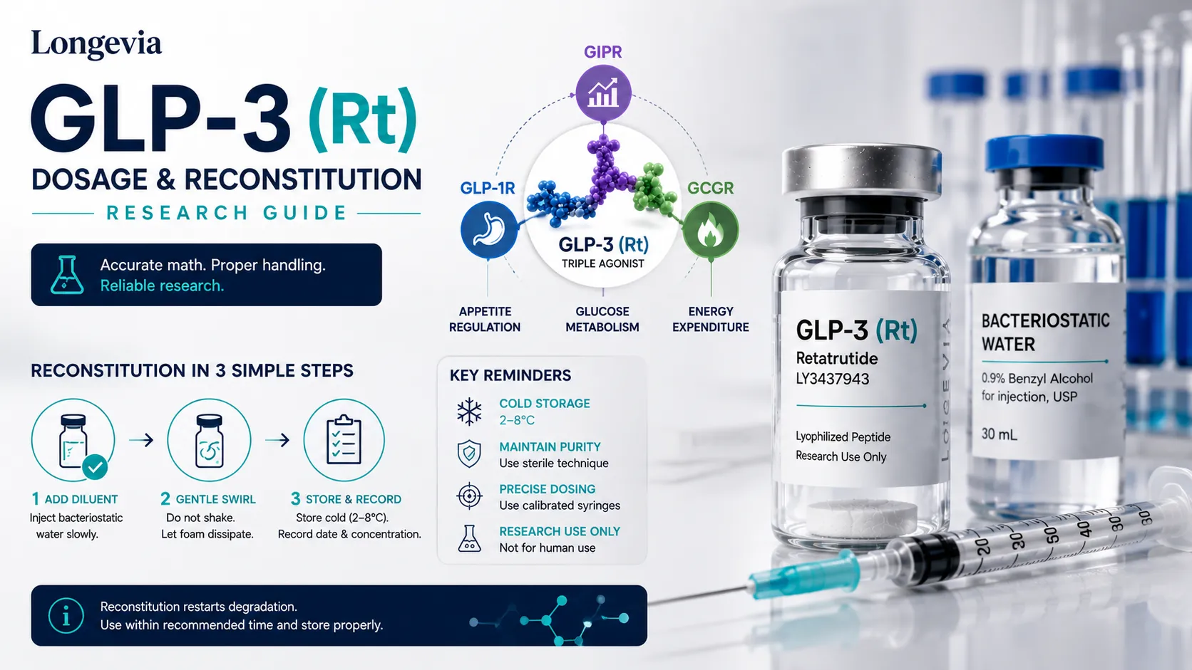
- Final concentration equals vial mass in milligrams divided by solvent volume in millilitres, because lyophilized powder adds negligible volume.
- A 10 mg vial reconstituted with 2 mL of bacteriostatic water yields a 5 mg per mL solution.
- Bacteriostatic water contains approximately 0.9 percent benzyl alcohol and suits multi-withdrawal containers; sterile water is single-use.
- Lyophilized material is the stable form and reconstituted solution is a consumable with a limited working life.
- Deamidation, hydrolysis, oxidation, and aggregation all accelerate once a peptide returns to aqueous solution.
- Repeated freeze-thaw cycling promotes aggregation, so single-use aliquots should be prepared at the point of reconstitution.
- Solvent should be introduced down the vial wall and the powder dissolved without shaking or vortexing.
- Lot number, concentration, solvent, and reconstitution date should be recorded on the vial label at the moment of reconstitution.
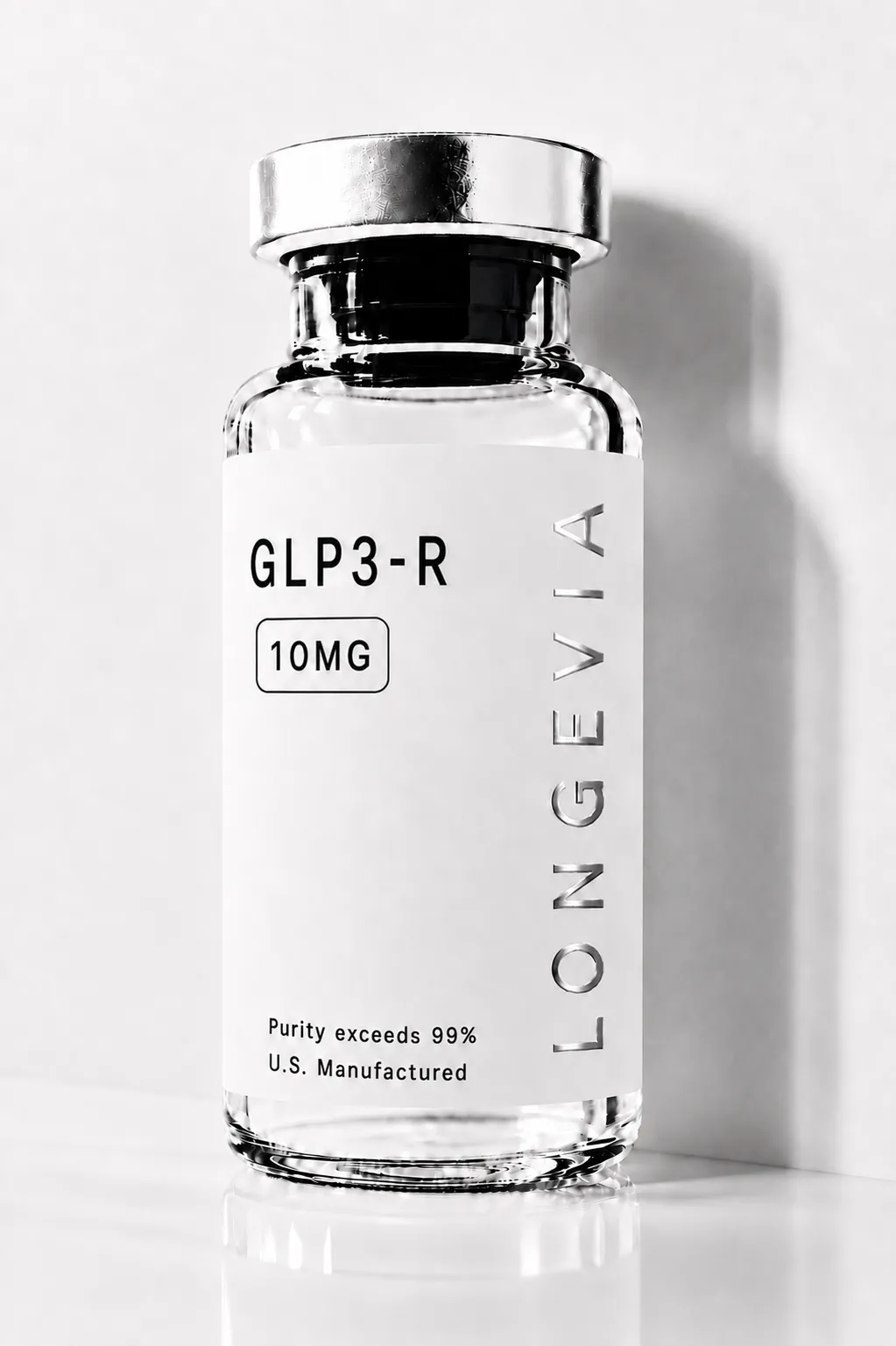
GLP-3 (Rt)
Explore GLP-3 (Rt) — a high-purity, COA-verified research peptide targeting GLP-1, GIP, and glucagon receptors. Lyophilized, batch-tested. Research use only.
GLP-3 (Rt)
Explore GLP-3 (Rt) — a high-purity, COA-verified research peptide targeting GLP-1, GIP, and glucagon receptors. Lyophilized, batch-tested. Research use only.
Reconstitution is the point at which a research compound stops being a stable, well-characterized solid and becomes a solution with a finite working life. For GLP-3 (Rt), the triple GIP, GLP-1, and glucagon receptor agonist also known as retatrutide (LY3437943), the arithmetic is straightforward but the handling is where most laboratory variability originates. This guide covers the reconstitution math that converts a lyophilized vial into a known concentration, the storage conditions that govern how long that concentration stays accurate, the degradation chemistry that explains why those conditions matter, and the protocol considerations specific to working with a three-receptor compound. All material discussed here is supplied for Research Use Only and is not for human consumption.
What Reconstitution Actually Does to a Lyophilized Peptide
Reconstitution rehydrates a freeze-dried peptide back into solution, and it also restarts every degradation pathway that lyophilization had suspended. This is the single most useful framing for the whole procedure. A lyophilized peptide sits in a low-water, low-mobility state in which the chemical reactions that break peptides down proceed extremely slowly. Adding solvent removes that protection.
Manning and colleagues catalogued the relevant chemistry in their review of protein and peptide pharmaceutical stability in Pharmaceutical Research, and the pathways are well established: hydrolysis of the peptide backbone, deamidation of asparagine and glutamine residues, oxidation of susceptible side chains, aggregation, and adsorption to container surfaces. All of these are accelerated by water, and most are further accelerated by temperature, unfavourable pH, agitation, and light exposure. Deamidation in particular is the most commonly observed chemical degradation route for peptides in aqueous solution.
The practical consequence for a research setting is that the concentration written on a label is only accurate for as long as the compound remains intact. A vial reconstituted six weeks ago and left at room temperature does not contain the concentration the arithmetic says it does, even though the volume has not changed. Degradation does not remove mass from the vial; it converts intact compound into related substances that a purity assay would separate but a volumetric calculation cannot see.
This is also why the acylated structure of retatrutide is relevant to handling. The molecule carries a fatty diacid moiety that supports albumin binding and an extended circulating half-life of approximately six days in the published pharmacokinetic work. That structural feature governs in vivo behaviour, not benchtop stability. Nothing about a long biological half-life implies a long shelf life in a reconstituted vial.
Lyophilized and reconstituted material are two different stability problems. Treat the freeze-dried vial as the stable form and the solution as a consumable with a defined working window, and record the reconstitution date on every vial.
Reconstitution Math: Bacteriostatic Water Volume to Final Concentration
Final concentration equals the mass of compound in the vial divided by the volume of solvent added, and nothing else in the calculation changes that relationship. The lyophilized powder contributes negligible volume, so the solvent volume added is effectively the final volume.
The formula is: concentration in mg per mL equals vial mass in mg divided by solvent volume in mL. The table below applies that formula to a 10 mg vial across common reconstitution volumes.
Bacteriostatic water added | Resulting concentration | Compound per 0.1 mL | Compound per 0.05 mL |
|---|---|---|---|
1.0 mL | 10 mg/mL | 1.0 mg | 0.5 mg |
2.0 mL | 5 mg/mL | 0.5 mg | 0.25 mg |
2.5 mL | 4 mg/mL | 0.4 mg | 0.2 mg |
4.0 mL | 2.5 mg/mL | 0.25 mg | 0.125 mg |
5.0 mL | 2 mg/mL | 0.2 mg | 0.1 mg |
For vial masses other than 10 mg, the same division applies. A 5 mg vial reconstituted with 2.0 mL yields 2.5 mg/mL. A 15 mg vial reconstituted with 3.0 mL yields 5 mg/mL. The choice of solvent volume is a trade-off: smaller volumes give higher concentrations and smaller withdrawal volumes, which increases the relative impact of pipetting error, while larger volumes give lower concentrations and larger, more accurately measurable withdrawal volumes at the cost of a shorter usable period per vial once opened.
Bacteriostatic water is water for injection containing approximately 0.9 percent benzyl alcohol as a preservative, which suppresses microbial growth in a multi-withdrawal container. Sterile water without preservative is single-use by design and offers no protection against contamination introduced during subsequent withdrawals. Some laboratory applications require preservative-free solvent because benzyl alcohol interferes with the assay or the model system, in which case single-use aliquoting becomes mandatory rather than optional.
For the general arithmetic behind concentration, dilution, and serial dilution calculations across any research compound, the dedicated calculation guide covers the full set of conversions.
Working Out Compound Mass Per Aliquot
The mass of compound in any withdrawn aliquot equals the concentration multiplied by the withdrawal volume, which is the only calculation needed once the vial concentration is fixed. This is the second half of the arithmetic and the half where unit errors most often occur, because concentrations are usually expressed in mg per mL while withdrawal volumes are usually measured in fractions of a mL.
Vial concentration | Withdrawal volume | Compound mass delivered | Equivalent in micrograms |
|---|---|---|---|
5 mg/mL | 0.02 mL | 0.1 mg | 100 mcg |
5 mg/mL | 0.10 mL | 0.5 mg | 500 mcg |
5 mg/mL | 0.20 mL | 1.0 mg | 1,000 mcg |
2 mg/mL | 0.05 mL | 0.1 mg | 100 mcg |
2 mg/mL | 0.25 mL | 0.5 mg | 500 mcg |
10 mg/mL | 0.05 mL | 0.5 mg | 500 mcg |
Two conventions prevent most calculation errors. First, standardize on one concentration across a study rather than reconstituting different vials to different concentrations, so that the volume-to-mass conversion becomes a single memorized ratio rather than a per-vial calculation. Second, record the vial mass, solvent volume, resulting concentration, and reconstitution date directly on the vial label at the moment of reconstitution rather than in a separate notebook, because the label travels with the material and the notebook does not.
Published human trial protocols for retatrutide used dose-escalation designs across a range of weekly amounts, reported in the phase 1b study by Urva and colleagues and the phase 2 trials published by Jastreboff and colleagues and Rosenstock and colleagues. Those figures describe the design of specific registered clinical protocols under investigator supervision. They do not constitute a protocol for research material, they carry no regulatory endorsement outside those trials, and they should not be mapped onto laboratory work or any other context.
Storage Conditions, Light Sensitivity, and Working Life
Lyophilized GLP-3 (Rt) is stored frozen and protected from light for long-term stability, while reconstituted solution is refrigerated, protected from light, and treated as having a limited working life. The table below summarizes the general conditions applied to research peptides of this class.
Material state | Temperature | Light | Practical working period |
|---|---|---|---|
Lyophilized, sealed, long term | -20 C or below | Protect from light | Longest stable state; see lot documentation |
Lyophilized, sealed, short term | 2 C to 8 C | Protect from light | Weeks to months |
Lyophilized, in transit | Ambient exposure tolerated briefly | Protect from light | Days; refrigerate on arrival |
Reconstituted, in use | 2 C to 8 C | Protect from light | Limited; verify against lot data |
Reconstituted, single-use aliquots | -20 C or below | Protect from light | Avoid repeated freeze-thaw cycles |
Three qualifications matter more than the table itself. First, compound-specific and lot-specific stability data always take precedence over general class guidance, which is one of the reasons a lot-specific Certificate of Analysis is more useful than a generic specification sheet. Second, general laboratory practice for peptides reconstituted in bacteriostatic water treats the refrigerated working window as a matter of weeks rather than months, but this is a practice convention rather than a compound-specific stability finding, and it should be treated as a working assumption to be verified rather than an established shelf life for this compound.
Third, freeze-thaw cycling is a distinct stress from storage temperature. Each cycle passes the solution through concentration and pH changes at the ice interface and promotes aggregation. Aliquoting into single-use volumes at the point of reconstitution removes the problem entirely and is worth the extra ten minutes on any material intended to last beyond a single session.
For a fuller treatment of post-reconstitution stability across compound classes, the dedicated stability guide covers the topic in depth.
Handling Protocol for Reconstitution
A reconstitution protocol has five steps, and the ordering of them is what protects both the compound and the sterility of the container. The sequence below reflects standard aseptic laboratory practice for lyophilized peptides.
- Equilibrate the sealed lyophilized vial to room temperature before opening. Introducing solvent into a cold vial promotes condensation, and opening a cold vial draws ambient moisture onto the powder.
- Disinfect both stoppers, the peptide vial and the solvent vial, and allow them to dry rather than wiping them dry.
- Draw the calculated solvent volume and introduce it down the inner wall of the vial rather than directly onto the powder cake. Direct high-velocity contact with the cake causes localized foaming and shear stress.
- Allow the powder to dissolve without agitation. Swirl gently if needed. Do not shake or vortex, because agitation generates air-liquid interfaces that drive aggregation and surface denaturation.
- Inspect the solution against a light source before use. A correctly reconstituted solution should be clear and free of visible particulates. Cloudiness, visible particles, or persistent foam are all reasons to stop and investigate rather than proceed.
Label immediately after step five with compound name, lot number, concentration, solvent used, and reconstitution date. Lot number in particular is the link back to the Certificate of Analysis for that specific batch, and once a vial is separated from its packaging that link is difficult to reconstruct.
Where a study will span several vials, recording the lot number alongside the reconstitution date allows any anomalous result to be traced back to a specific batch rather than attributed to the compound in general. Material from a single lot should be preferred within a single experimental series wherever supply allows.
GLP-3 (Rt) is supplied by Longevia Research strictly for Research Use Only. It is not a medicine, it has not been approved by the FDA, the EMA, or any equivalent authority for any indication, and it is not intended for human or veterinary consumption, diagnostic use, or therapeutic use. Nothing in this guide describes or implies any permitted use in humans.
Research Protocol Considerations Specific to a Triple Agonist
Working with a triple receptor agonist introduces experimental design considerations that a single-receptor compound does not, because any observed effect has three possible mechanistic origins rather than one. This is a study design problem rather than a handling problem, but it shapes how material is allocated and therefore how vials are reconstituted.
Retatrutide activates the GIP, GLP-1, and glucagon receptors from a single molecule, and Coskun and colleagues reported that its potency is not evenly distributed across those three targets. Attributing an observed readout to a specific receptor arm therefore requires either selective antagonists, receptor-deficient model systems, or dose-ranging designs that exploit the differing potency thresholds across the three receptors. Each of these approaches consumes more material than a single-arm design, which is a practical argument for reconstituting to a concentration that supports the full dose range in a single vial rather than across several.
Concentration choice interacts directly with this. A dose-ranging design spanning two orders of magnitude is much easier to execute accurately from a single stock with serial dilutions than from multiple independently reconstituted vials, because serial dilution propagates one weighing and one volumetric measurement rather than several. Preparing a single high-concentration stock and diluting from it also keeps the number of container-surface adsorption events per aliquot consistent across conditions.
Vehicle controls deserve specific attention when bacteriostatic water is the solvent. Benzyl alcohol is biologically active at sufficient concentration, so a vehicle control matched for benzyl alcohol content, not just for volume, is the appropriate control in any system where solvent effects are plausible.
The same logic applies to any co-solvent used to aid dissolution, which should appear in the control arm at matched concentration rather than being assumed inert. Solvent-matched controls are cheap to run and expensive to omit, because a solvent effect discovered after data collection cannot be separated from a compound effect retrospectively.
For the receptor pharmacology underlying all of this, including the relative potency data and the signaling characteristics of each arm, the mechanism guide covers the primary literature.
Verifying the Material Before You Reconstitute It
Verification belongs before reconstitution rather than after, because once solvent is added the material cannot be returned to its stable state and any identity or purity problem has been carried into the experiment. Three checks take a few minutes and eliminate most of the risk.
The first is matching the lot number on the vial to a lot-specific Certificate of Analysis rather than to a generic product specification. A generic specification describes what a product is supposed to be. A lot-specific COA describes what a particular batch actually tested as, which is the only document that says anything about the vial in hand. Longevia Research publishes lot-specific COAs in its COA Library for this reason.
The second is confirming that the COA reports both a quantitative purity determination by HPLC and an identity confirmation by LC-MS. HPLC separates the target compound from related substances and reports purity as a percentage of the chromatographic profile. LC-MS confirms that the main peak has the expected molecular mass, which is what distinguishes the correct compound from a different compound of similar retention behaviour. Purity without identity is an incomplete result, because a highly pure sample of the wrong molecule still reports a high purity figure.
The third is a physical inspection of the lyophilized cake before any solvent is added. A well-lyophilized peptide cake is uniform and intact. A collapsed, melted, discoloured, or visibly wet cake indicates a temperature excursion somewhere in the supply chain, and no reconstitution procedure recovers material that has already been compromised.
USP General Chapter 1503, which describes quality attributes for synthetic peptide drug substances, provides the framework these analytical conventions follow, including identity testing by orthogonal methods and the characterization of peptide-related impurities. Research suppliers are not operating under pharmacopoeial drug substance requirements, but the analytical logic of orthogonal identity confirmation plus quantitative purity determination is the correct standard to expect from documentation regardless of the regulatory context.
Frequently Asked Questions
- Manning MC, Chou DK, Murphy BM, Payne RW, Katayama DS. Stability of protein pharmaceuticals: an update. Pharm Res. 2010;27(4):544-575.
- Coskun T, Urva S, Roell WC, et al. LY3437943, a novel triple glucagon, GIP, and GLP-1 receptor agonist for glycemic control and weight loss: from discovery to clinical proof of concept. Cell Metab. 2022;34(9):1234-1247.e9.
- Urva S, Coskun T, Loh MT, et al. LY3437943, a novel triple GIP, GLP-1, and glucagon receptor agonist in people with type 2 diabetes: a phase 1b, multicentre, double-blind, placebo-controlled, randomised, multiple-ascending dose trial. Lancet. 2022;400(10366):1869-1881.
- United States Pharmacopeia. General Chapter 1503, Quality Attributes of Synthetic Peptide Drug Substances. USP-NF. Rockville, MD.
- Jastreboff AM, Kaplan LM, Frias JP, et al. Triple-Hormone-Receptor Agonist Retatrutide for Obesity - A Phase 2 Trial. N Engl J Med. 2023;389(6):514-526.